Surgical Canine Capabilities. Before making the puncture the venipuncturist can lay the thumb of the hand that is holding the leg adjacent to the vein to reduce vein movement when Placing a catheter in the cephalic or saphenous vein.
2
Clip the haired skin overlying the site for IV catheterization.
Canine cephalic vein. Tape to secure the IV catheter in place. Replacement Canine Leg Cephalic Vein. Please allow for a production time of up to 10 business days before your item ships.
The cephalic vein is a superficial vein of the upper limb and its one of the two main veins of the arm. If dog is unconscious can use a tourniquet. The external jugular vein then divides into a dorsal branch maxillary vein and ventral vein lingofacial vein at the caudal border of the dog mandibular gland.
Gastrotomy with foreign body removal. The cephalic vein can have a tortuous course in the brachicephalic breeds making location and fixation of the vein for catheterization difficult. Watch how to place an IV catheter in the cephalic vein of a dog.
Cephalic vein saphenous vein or jugular vein caudo-dorsal neck or thorax uncommon thorax or abdomen cattle neck muscle jugular vein tail vein neck or shoulder NA thorax or neck goatsheep neck muscle jugular vein skin below elbow NA thorax or neck pig base of ear ear vein anterior vena cava jugular vein. Place the animal in sternal or lateral recumbency. To collect blood from a peripheral vein introduce the needle into the occluded vessel as far distally as possible.
Locate the cephalic vein. Once blood collected let off vein BEFORE the needle removed still hold onto leg then cover puncture site when needle out. The cephalic vein usually receives three tributaries in the antebrachium.
To facilitate venipuncture of this vessel follow the same steps as for the cephalic vein. 3 UK-14304 is a partial agonist at alpha 1-adrenoceptors. Intestinal resection with anastomosis.
This vein gives off cephalic superficial cervical and omobrachial veins on its caudal-cranial sequences. Characteristics of normal canine cephalic veins were as follows. Includes equipment preparation and bandaging technique.
The cephalic jugular femoral and medial saphenous veins are used for feline venipunctures. Using sterile techniques a superficial transverse cut down starting from the midmedial antebrachium to lateral is performed. In this case I have put a tourniquet on the elbow so that the vein fills with blood and is easier to see.
2 according to the original definition of spare receptors there is no alpha 1-adrenoceptor reserve in canine cephalic vein. The image below shows the cephalic vein. Occlude the cephalic vein.
1 the canine cephalic vein is a suitable preparation to study postsynaptic alpha 1- and alpha 2-adrenoceptors. 40 clipper blade iii. The cephalic vein arises here from the radial vein as the main conduit of venous blood from the palmar surface of the paw.
Let off vein once IV catheter is in and move to hold off vein at the proximal end of catheter to prevent blood from leaking. Extend the arm at the elbow. Enterotomy with foreign body removal.
The site is easily accessible for interference by the patient - catheter must be secured adequately and interference prevented eg using an Elizabethan collar or muzzle. Dogs can either be restrained manually or using a sling. The accessory cephalic vein is an alternative venipuncture site in medium to large dogs.
The most frequently used sites for canine blood collection are the cephalic jugular and lateral saphenous veins. Lateral saphenous vein ii. Again the cephalic vein lies at the medial part of the lateral pectoral groove.
Dorsomedial aspect at the level of carpus distal to the cephalic vein. The cephalic vein is located on the front of the foreleg the dorsal surface. Smooth and thin wall complete compressibility no flow disturbances no filling defects smooth flow contours and unidirectional non-pulsatile flow with no turbulence.
Prepare the site i. This is a common site of insertion in lizards and is located running from medial to lateral in a proximal to distal direction across the antebrachium Fig. If dog is conscious handler places thumb over the cephalic vein and heel of the hand under the dogs elbow.
Hold off vein and position it dorsal on the foreleg-make sure comfortable for you and the dog. Now follow the photos with instructions below. Occlude the cephalic vein.
Your assistant should maintain the animal in this position. Our simulated cephalic vein is softer than typical latex tubing to enhance Train on venipuncture and vascular access skills with SurgiReals Canine Leg Vascular Access Simulator. OVH replaceable Cystotomy with stone removal.
Cephalic vein on the medial side of the limb at a location distal to the junction of the cephalic and accessory cephalic veins. Restraint for a Canine Cephalic Vein Blood Draw - YouTube. The smaller radial vein continues proximally in the antebrachial muscles.
Replacement cephalic vein for the Canine Vascular Access Simulator. More in this package. Made of high quality durable silicones the Canine Leg is designed specifically for blood collection and catheter insertion of a 22 gauge or smaller.
The vein runs under the skin between the carpal wrist joint and the elbow. Quantity must be 1 or more. Accessory Cephalic Vein i.
It is concluded that. Its name derives from cephalic meaning head as the vein runs up to the shoulderThe superficial venous network is the source of blood for most blood tests and is the easiest place to access venous blood.
Before making the puncture the venipuncturist can lay the thumb of the hand that is holding the leg adjacent to the vein to reduce vein movement when Placing a catheter in the cephalic or saphenous vein. Clip the haired skin overlying the site for IV catheterization.

Iv Injection And Blood Collection Flashcards Quizlet
Hold off vein and position it dorsal on the foreleg-make sure comfortable for you and the dog Once blood collected let off vein BEFORE the needle removed still hold onto leg then cover puncture site when needle out.
Cephalic vein dog. Cannulation of the cephalic basilic or other. The vein runs under the skin between the carpal wrist joint and the elbow. Dorsomedial aspect at the level of carpus distal to the cephalic vein.
The patient with the ligated radial artery had a confirmed patent ulnar artery pre- and postoperatively. Intestinal resection with anastomosis. Surgical Canine Capabilities.
Prepare the site i. Cephalic Venipuncture To collect blood from the cephalic vein which is located on the cranial aspect of the foreleg the animal is restrained in sternal recumbency or in a standing position. Enterotomy with foreign body removal.
The axillobrachial vein is ligated and divided as it branches from the cephalic vein. Your assistant should maintain the animal in this position. The cephalic vein is a superficial vein of the upper limb and its one of the two main veins of the arm.
Again the cephalic vein lies at the medial part of the lateral pectoral groove. Cephalica accessoria at the. Its name derives from cephalic meaning head as the vein runs up to the shoulder.
The superficial venous network is the source of blood for most blood tests and is the easiest place to access venous blood. Blood collection from cephalic vein is bit easier as compare to others as restraint of the pet is convenient and this vein locates easily. Shave a wide margin to maintain a sterile field Ideally make sure to have a clean looking symmetrical shave job unless its a life-threatening emergency.
Because of its size location and ease of compressibility it is the favored site for venipuncture in the dog. The image below shows the cephalic vein. The acromion portion of the deltoideus muscle is transected at its insertion on the deltoid tuberosity of the humerus and is reflected proximally.
To facilitate venipuncture of this vessel follow the same steps as for the cephalic vein. The antebrachial part of the cephalic vein is augmented by receiving the accessory cephalic vein v. How to Place an IV Catheter in the Cephalic Vein Dog.
Along the outer side of the arm superficial to the deep fasciaruns the cephalic vein. In the two patients with ligations only the affected vessels were the radial artery and cephalic vein. Additionally which veins are used for cannulation.
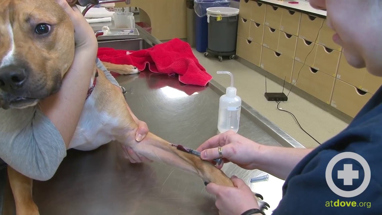
The study was performed to investigate the difference in the concentration of several haematological and clinical chemical blood components in blood obtained from the cephalic and external jugular veins in 23 dogs. To start have the restrainer gently holding the patient behind the elbow to secure the limb. The accessory cephalic vein is an alternative venipuncture site in medium to large dogs.
Gastrotomy with foreign body removal. Small dogs may be picked up and held in the restrainers arms with a foreleg extended. The vein runs under the skin between the carpal wrist joint and the elbow.
Includes equipment preparation and bandaging technique. This vein gives off cephalic superficial cervical and omobrachial veins on its caudal-cranial sequences. Cephalic vein on the medial side of the limb at a location distal to the junction of the cephalic and accessory cephalic veins.
In the remaining two bypass-only cases a reversed femoral vein 113 and reversed cephalic vein 113 were used. The image below shows the cephalic vein. Watch how to place an IV catheter in the cephalic vein of a dog.
The cephalic vein is often the most common and easiest location to use for catheterization. The cephalic vein is located on the front of the foreleg the dorsal surface. The external jugular vein then divides into a dorsal branch maxillary vein and ventral vein lingofacial vein at the caudal border of the dog mandibular gland.
The cephalic vein is located on the front of the foreleg the dorsal surface. If the right vein is to be tapped or catheterized the assistant should stand on the left side of the animal and place the left arm or hand under the animals chin to immobilize the head and neck. Place the animal in sternal or lateral recumbency.
How to Do a Canine Cephalic Vein Blood Collection - YouTube. OVH replaceable Cystotomy with stone removal. Small dogs may be picked up and held in a standing position.
A total of 17 laboratory tests were analysed and except for two clinical chemical bl. Accessory Cephalic Vein i. Above this vein is found in the groove betweenthe pectoralis major and the deltoid and after piercing the costo-coracoid membrane passes across the first part of.
To restrain a dog or cat for venipuncture of the cephalic vein place the dog or cat on the table sitting or in sternal recumbency. Similarly you may ask where is the cephalic vein on a dog. Restraint for a Canine Cephalic Vein Blood Draw - YouTube.
Dogs can either be restrained manually or using a sling. Lateral saphenous vein ii. The cleidobrachialis muscle is transected at its insertion on the distal aspect of the humeral crest.
To view the veinextended the elbow of the pet and put the index finger on the front of the leg above elbow joint and thumb at.